

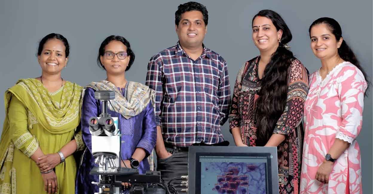
A research team from the National Institute of Technology Calicut has developed an innovative, AI-assisted smart microscopy system designed to revolutionise medical image analysis. By integrating advanced artificial intelligence with traditional optical microscopes, this breakthrough device allows pathologists to diagnose illnesses with unprecedented speed and precision.
An intelligent upgrade for traditional microscopes
The newly developed system serves as an easy-to-integrate upgrade for existing microscopes, turning conventional laboratory equipment into highly intelligent diagnostics setups. It works by analysing slide images in real time, automatically highlighting abnormal cells, micro-anomalies, and other critical pathology markers for clinicians. This immediate screening drastically reduces eye strain and cognitive fatigue for laboratory specialists, who traditionally spend long hours staring into eyepieces, while accelerating report turnaround times.
Revolutionising rural healthcare with offline edge AI
A standout feature of this end-to-end device is its edge-computing design, allowing all AI processing to occur directly on the device itself. Because it does not rely on an active internet connection or cloud infrastructure, the system ensures optimal data privacy and operates perfectly in resource-limited environments. This self-contained structure makes it highly viable for primary health centres, community clinics, and remote rural hospitals that lack advanced computing facilities.
From cancer detection to wider pathology
While initially configured and validated for urine cytology tests—specifically for the detection of bladder cancer—the underlying technology is highly adaptable. The researchers plan to expand its applications to cover a broad spectrum of cytology and pathology analyses, including haematology, liver health screens, and tissue pathology. This synergy of medical expertise and engineering represents a major leap forward in diagnostic technology.
Collaborative genius and market launch
This cutting-edge technology was developed by a collaborative team comprising Computer Science faculty and researchers from NIT Calicut, working alongside pathology experts from Government Medical College Calicut and KMCT Medical College. The innovation is scheduled to be commercialised and marketed through Healforge AI Private Limited, an enterprise incubated at NIT Calicut, bringing this clinic-ready AI microscope to diagnostic labs globally.
National funding and the visionary team
The developmental research for this invention was funded by the Ministry of Education, Government of India, under the Scheme for Promotion of Academic and Research Collaboration (SPARC) initiative.
The key innovators behind this development include:
- Aarathi Menon, Research Scholar, Department of Computer Science and Engineering, NIT Calicut
- Dr Jayaraj PB, Associate Professor, Department of Computer Science and Engineering, NIT Calicut
- Dr Pournami PN, Associate Professor, Department of Computer Science and Engineering, NIT Calicut
- Dr Simi S, Assistant Professor, Department of Pathology, Government Medical College Calicut
- Dr Swathi Shankar, Assistant Professor, Department of Pathology, KMCT Medical College Calicut